Hysteroscopy
A Clearer Look Inside the Uterus for Better Fertility Care
Hysteroscopy is a minimally invasive procedure that allows a fertility specialist to examine the inside of the uterus using a thin, lighted instrument called a hysteroscope.
It provides a direct view of the uterine cavity and can help identify certain conditions that may affect menstrual health, implantation, or pregnancy. Under the expert clinical guidance of Dr. Devkriti Tiwari Dhirawani, diagnostic and operative hysteroscopy restores the uterine environment to its healthiest state for natural conception or assisted reproductive cycles (IVF / IUI).
Direct View
HD Cavity InspectionMinimally Invasive
Zero Abdominal CutsDaycare Care
Rapid Same-Day HomeFertility Care
Implantation Ready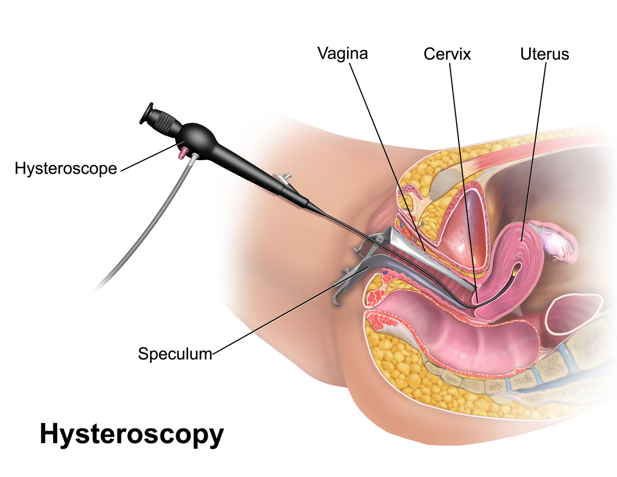
What Is Hysteroscopy?
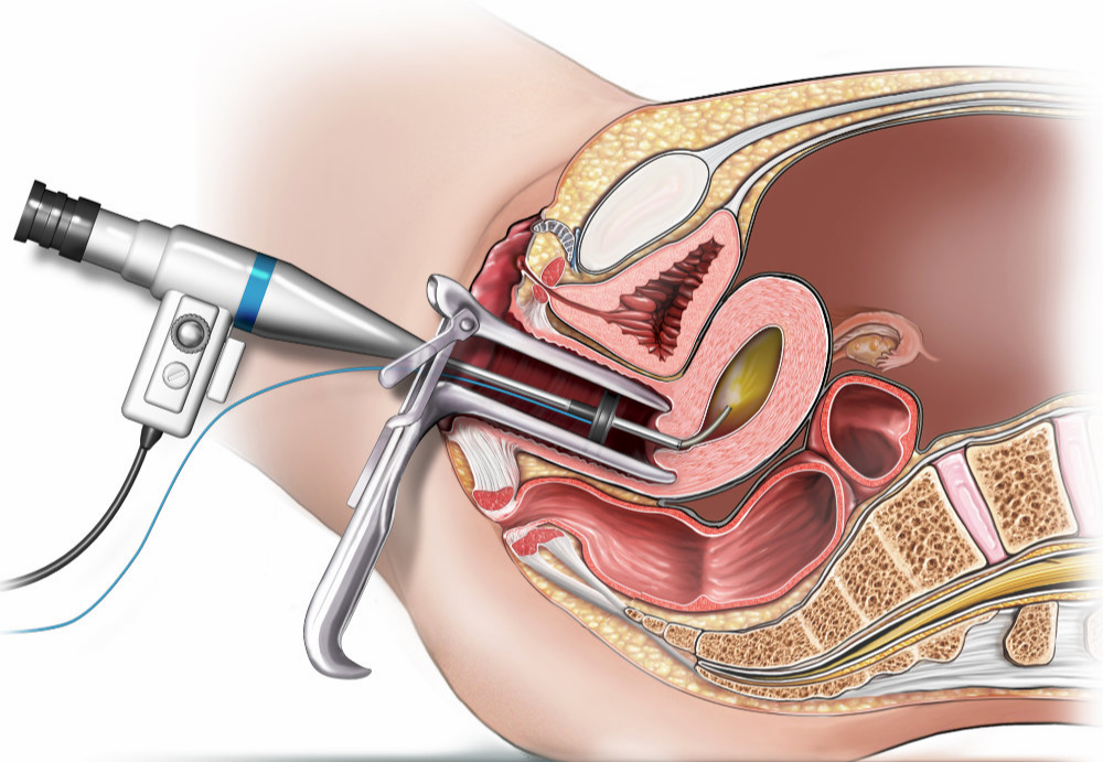
Hysteroscopy is a procedure used to carefully examine the uterine cavity. A slender camera is gently guided through the natural passage of the vagina and cervix, allowing the doctor to view the inside of the uterus on a high-definition monitor screen.
If a problem is identified, certain conditions can also be treated during the same procedure (known as operative hysteroscopy), depending on the patient's individual clinical needs.
Key Clinical Advantages:
- Direct Optical Clarity: Eliminates guesswork from 2D ultrasounds by providing direct visualization.
- Single-Session Treatment: Enables diagnosis and micro-surgical correction in a single sitting.
- No External Scarring: Performed completely through natural channels without incisions.
- Optimizes Endometrium: Prepares the uterine lining for healthy embryo attachment and full-term growth.
Why Might Hysteroscopy Be Recommended?
Your fertility specialist may recommend hysteroscopy when there is a possibility of an abnormality inside the uterus. It helps in thoroughly evaluating and addressing:
Uterine Polyps
Small fleshy growths inside the uterine cavity that may interfere with regular menstrual bleeding or embryo implantation.
Submucosal Fibroids
Certain fibroids that develop within or close to the inner uterine cavity lining, distorting cavity contour and requiring evaluation.
Uterine Adhesions
Intrauterine scar bands (Asherman's syndrome) that can develop after past procedures or infections, restricting healthy endometrium.
Abnormal Bleeding
Helps investigate and diagnose underlying physical causes of unexplained, heavy, or irregular uterine bleeding episodes.
Recurrent Pregnancy Loss
A thorough evaluation of the uterine cavity architecture in selected cases of repeated miscarriages or recurrent implantation failures.
Fertility Evaluation
In patients preparing for IVF or fertility treatments, examining the cavity provides decisive confirmation that the uterus is ready for implantation.
How Can Hysteroscopy Help in Fertility Care?
The uterus plays a pivotal role in embryo implantation and pregnancy. An undetected abnormality within the uterine cavity—such as a small polyp, a partial septum, or scar tissue—may act as a physical barrier or create localized inflammation that prevents an embryo from implanting successfully.
Hysteroscopy gives the fertility specialist a direct, panoramic view of the uterine cavity rather than relying only on indirect imaging tests. If a treatable abnormality is discovered, corrective micro-treatment can be planned and executed according to the patient's individual condition.
The Goal is Simple:
Diagnostic vs. Operative Hysteroscopy
Explore the two complementary modalities of hysteroscopic care provided at our centre:
Diagnostic Hysteroscopy (Visual Assessment)
Primarily focused on clear visual inspection of the endocervical canal, uterine cavity, and bilateral fallopian tubal ostia. Conducted to investigate abnormal bleeding, check cavity architecture prior to IVF, or confirm findings from previous ultrasound / HSG scans.
- Duration: 10 to 15 minutes
- Anesthesia: Local cervical block or light mild sedation
- Recovery: Discharge within 1 to 2 hours (Outpatient)
- Primary Goal: HD visualization & accurate anatomical confirmation
Common Indications for Diagnostic Hysteroscopy:
Operative Hysteroscopy (Targeted Micro-Correction)
Combines visualization with specialized miniature surgical instruments passed through operative hysteroscopic channels. Enables real-time correction of anatomical defects without any incisions on the abdomen.
- Duration: 20 to 45 minutes
- Anesthesia: Monitored intravenous sedation or gentle spinal
- Recovery: Same-day discharge (Daycare procedure)
- Primary Goal: Micro-surgical excision, reshaping, and restoration
Conditions Corrected Operatively:
What Happens During Hysteroscopy?
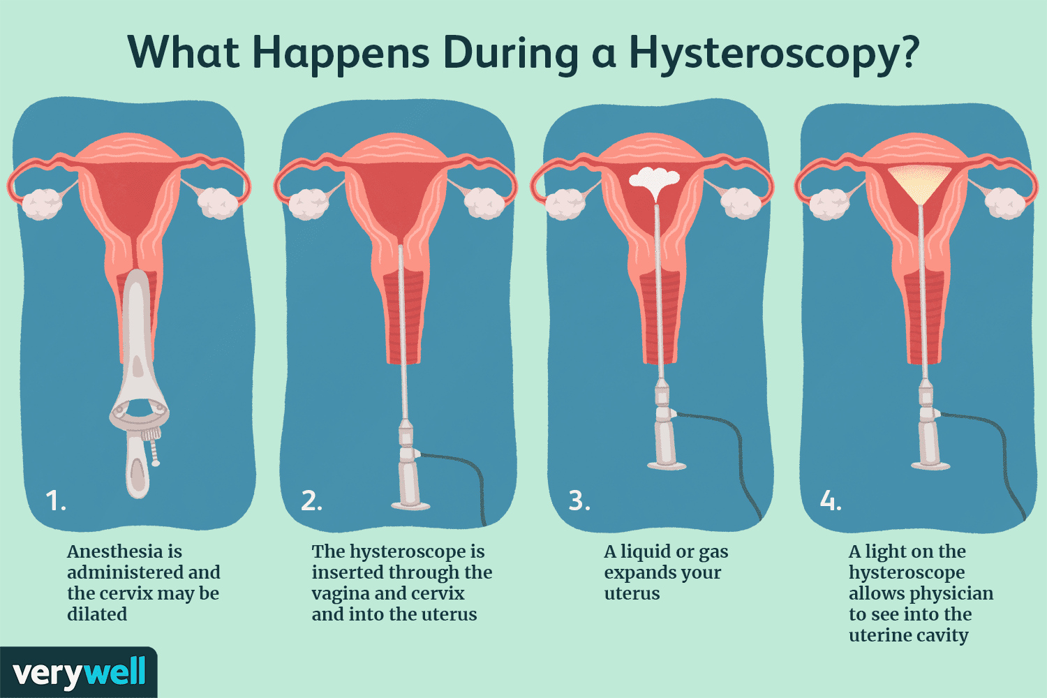
A clear, five-stage clinical progression ensuring patient comfort, safety, and thorough diagnosis:
Initial Evaluation
Your fertility specialist evaluates your symptoms, medical history, and previous ultrasound scans to determine whether hysteroscopy is appropriate.
Gentle Insertion
A thin, lubricated hysteroscope is carefully introduced through the vagina and cervix into the uterus, requiring no surgical incisions.
Clear Visualization
A small camera provides a magnified, high-resolution view of the uterine cavity, allowing the doctor to examine its internal lining and structure.
Treatment, If Required
If a suitable abnormality is identified (such as a polyp or intrauterine adhesion), corrective micro-surgical procedures can be performed during the same session.
Follow-Up & Next Steps
Your doctor discusses the findings with you in detail, sharing digital endoscopic images and recommending the appropriate next step based on your fertility goals.
Benefits of Hysteroscopy
Why hysteroscopy is considered the clinical gold standard for uterine cavity evaluation:
Direct Visualization
Allows the doctor to see the uterine cavity directly in real time under true magnification.
Minimally Invasive
Uses a thin instrument through natural passages and does not require a large surgical incision.
Detects Abnormalities
Assists in detecting conditions such as polyps, certain fibroids, septums, and adhesions.
Treatment May Be Possible
Some abnormalities can be treated during hysteroscopy when clinically appropriate, avoiding a second procedure.
Supports Fertility Planning
Findings from the procedure can help your fertility specialist decide on the most effective treatment protocol.
Is Hysteroscopy Right for Everyone?
Not every patient undergoing fertility treatment needs hysteroscopy.
Your doctor may recommend it based on your specific symptoms, medical history, previous ultrasound investigations, and fertility treatment history. A personalized evaluation is important before deciding whether the procedure is appropriate for your unique situation.
Frequently Asked Questions About Hysteroscopy
Medically verified answers to common patient questions regarding hysteroscopy and uterine health: